
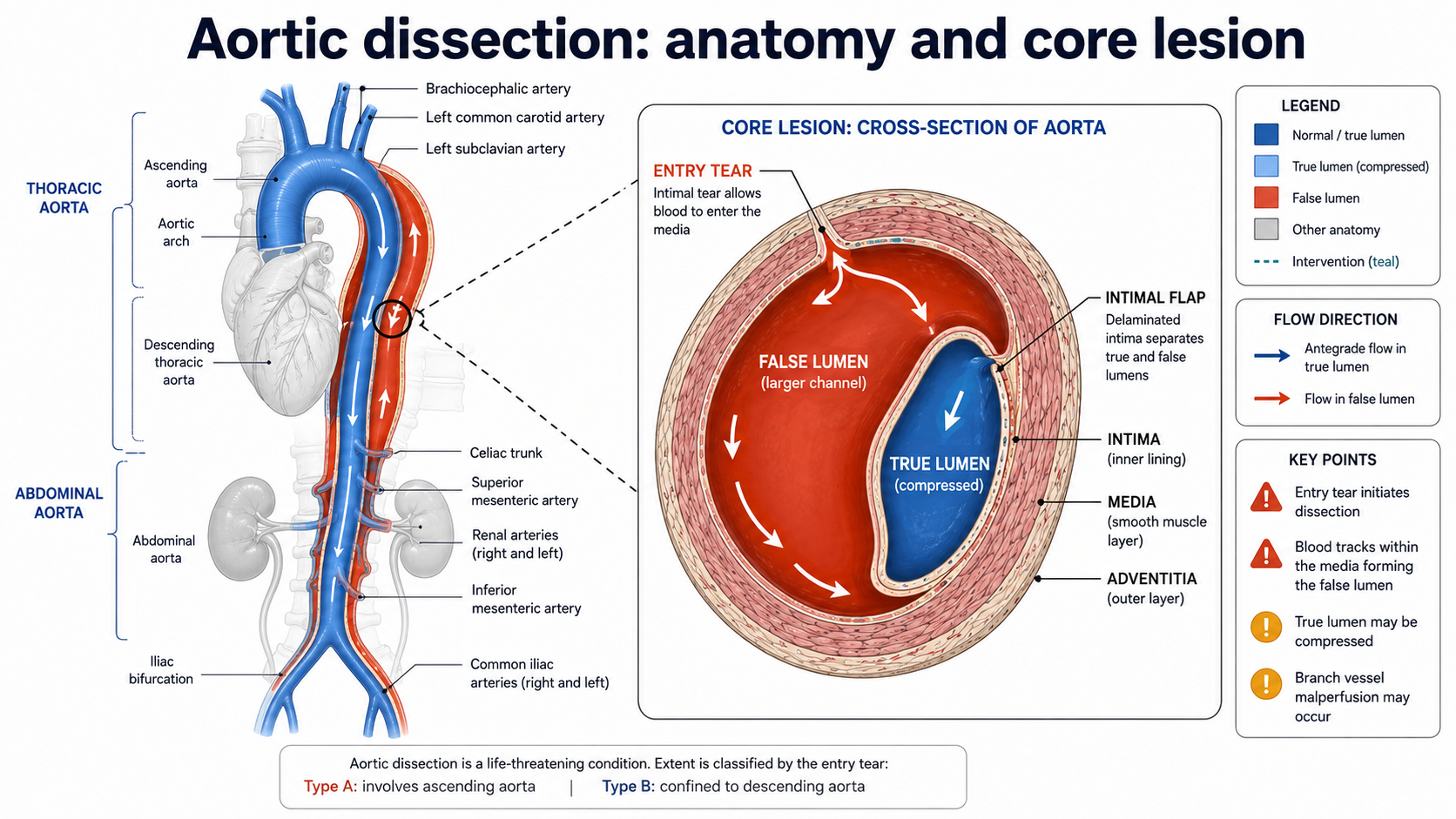
Aortic dissection is the archetypal disease in which anatomy, time, haemodynamics, imaging, surgical judgement and systems of care intersect. It belongs to the spectrum of acute aortic syndromes, together with intramural haematoma and penetrating atherosclerotic ulcer, but retains a distinct clinical identity because the essential lesion is propagation of blood within the aortic media after disruption of the intima or of the medial layers. The result is a double-channel aorta, with a true lumen and a false lumen separated by an intimal flap. The false lumen may re-enter distally, remain pressurized without re-entry, thrombose partially or completely, compress the true lumen, occlude branch vessels, rupture externally, or persist as the substrate for late aneurysmal degeneration. These anatomical possibilities explain why aortic dissection is not a single therapeutic problem but a family of time-sensitive syndromes in which the treatment target is determined by the affected aortic segment, the entry tear, the presence of malperfusion, rupture risk, patient comorbidity and centre expertise.1–4
Historical evolution: from pathological curiosity to systems-based emergency
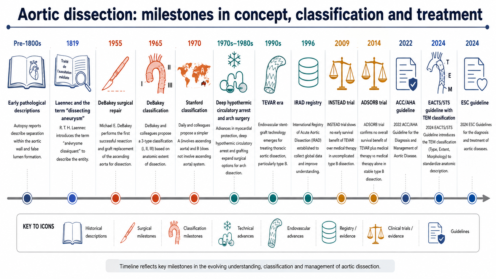
The intellectual history of aortic dissection begins in pathology and autopsy, not in imaging or intervention. Early descriptions of fatal aortic rupture and dissecting aneurysm predated modern cardiovascular surgery by centuries. The older term “dissecting aneurysm”, popularized in the Laennec era, reflected the observation of blood dissecting within the wall but also embedded a conceptual confusion: many dissections are not aneurysms at presentation, and many aneurysms do not dissect. This terminological residue still matters because it can obscure the immediate pathophysiological distinction between acute medial separation and chronic aneurysmal dilation.
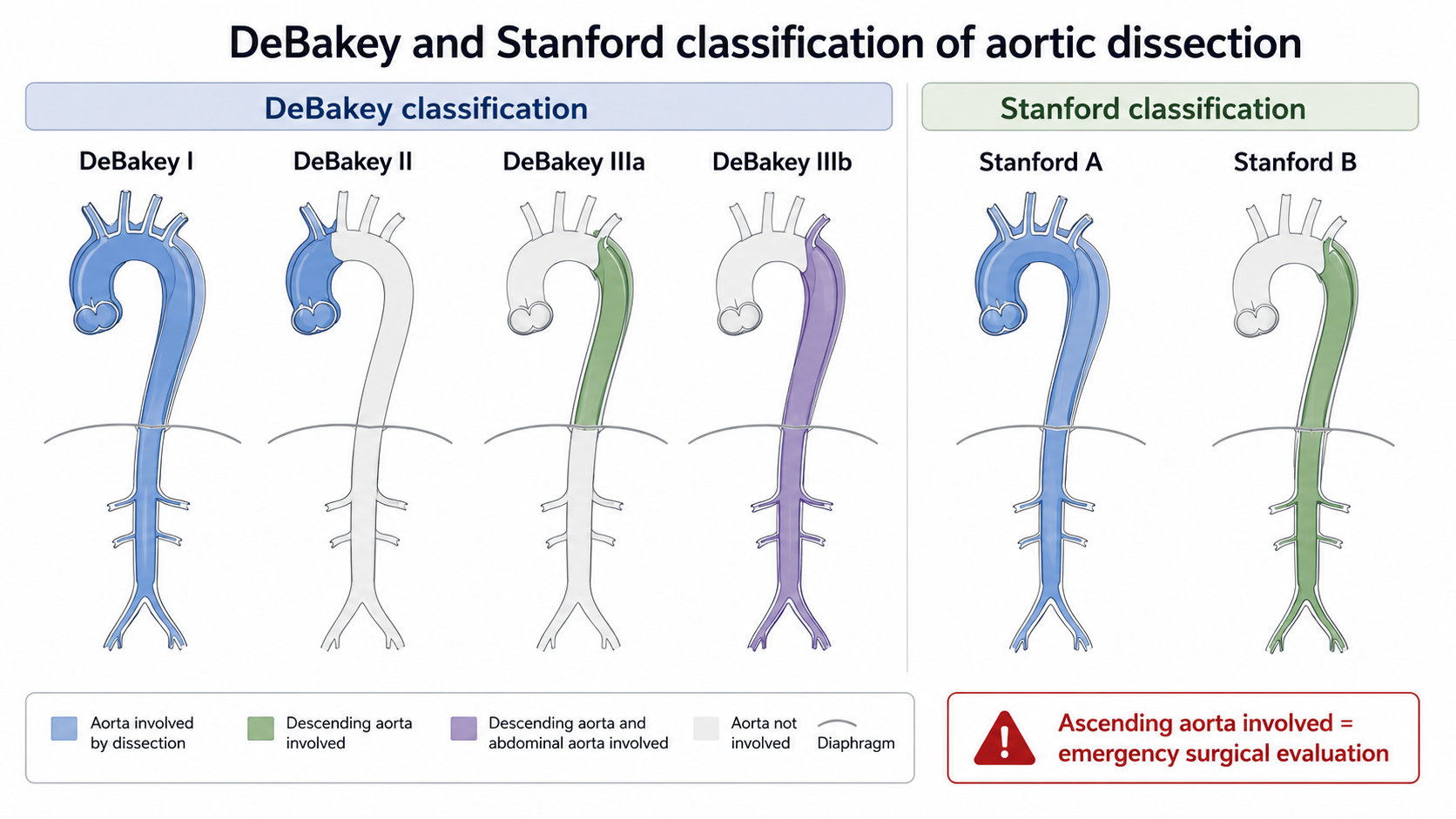
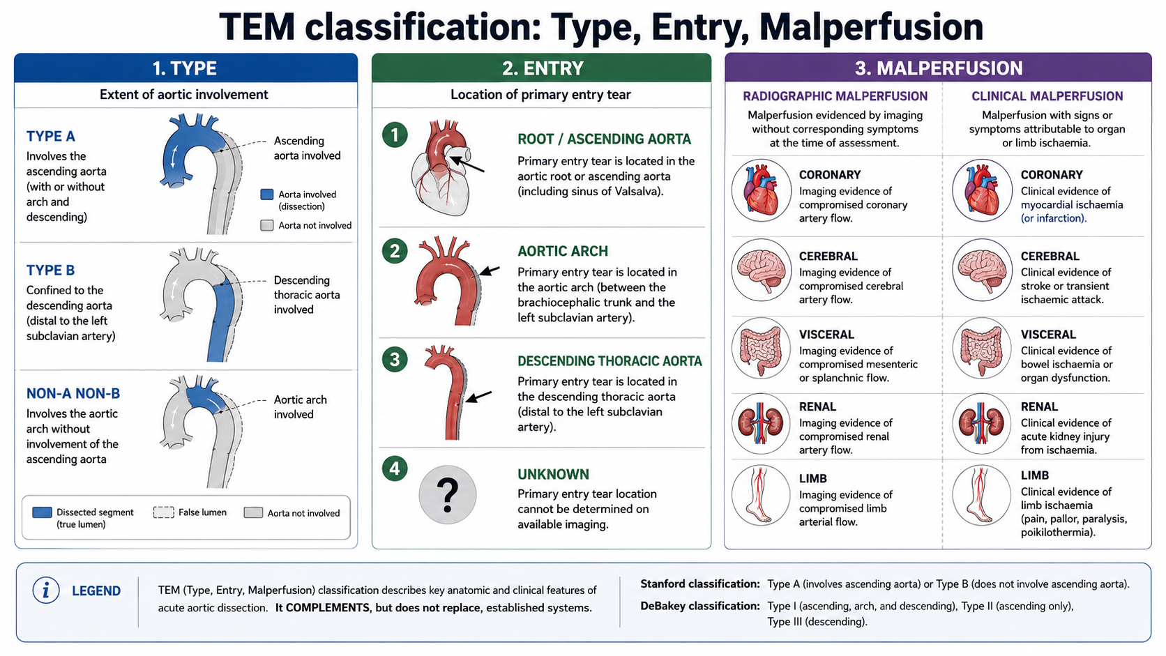
Modern operative treatment began in the mid-twentieth century. The first successful surgical repairs reported by DeBakey, Cooley and Creech established that dissection was not uniformly fatal once the entry tear and diseased aortic segment could be surgically controlled.6 DeBakey classification then organized the disease by the site and extent of dissection: type I involving the ascending aorta, arch and descending aorta; type II confined to the ascending aorta; and type III beginning in the descending thoracic aorta, with IIIa and IIIb reflecting distal extension. Stanford classification, introduced by Daily, Shumway and colleagues, deliberately simplified the decision problem: type A if the ascending aorta is involved, type B if it is not.7 The move from DeBakey to Stanford was not merely semantic; it converted anatomical description into a management algorithm.
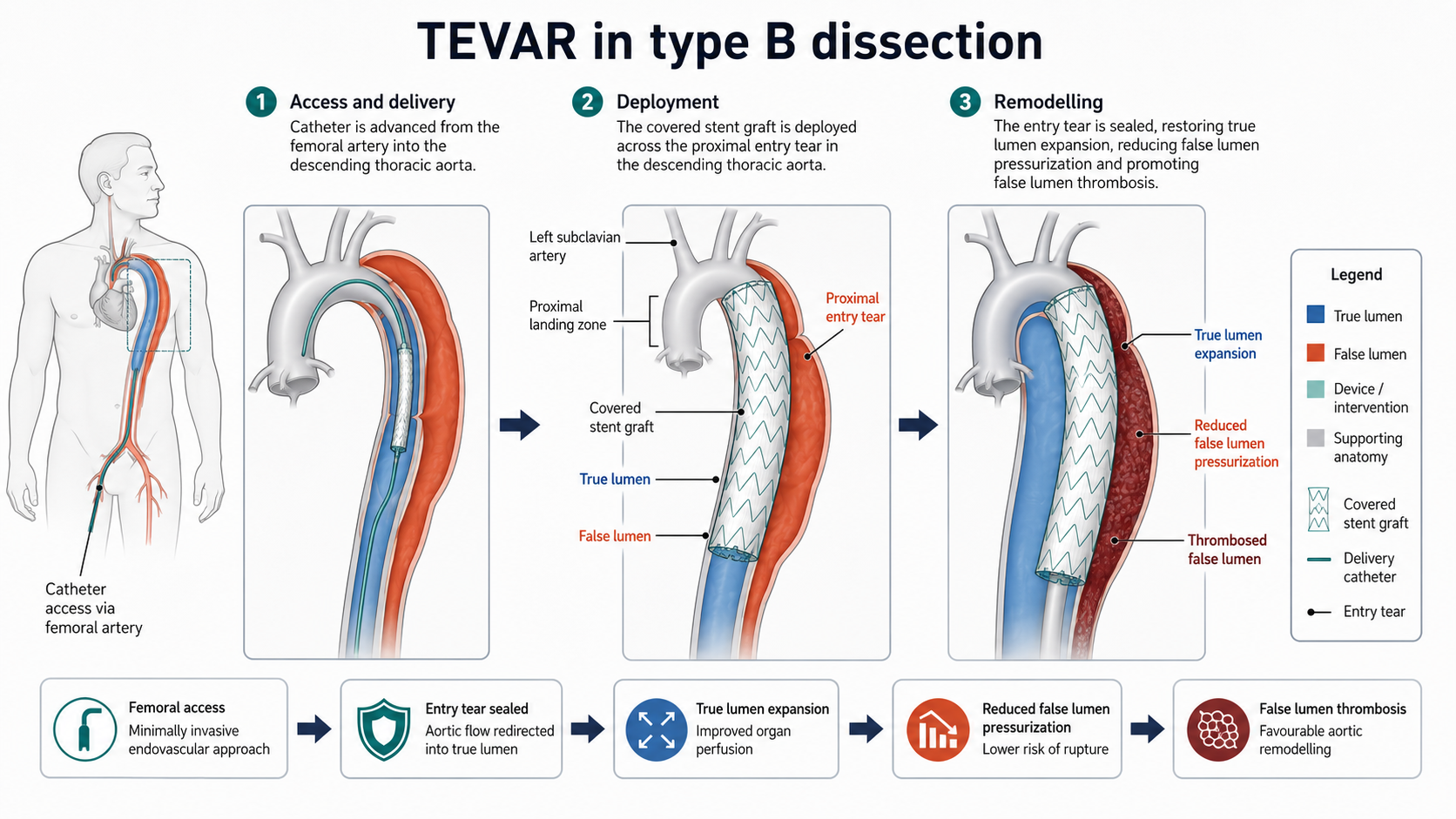
The subsequent evolution of the field has occurred through three major transitions. First, cardiothoracic surgery moved from heroic replacement of the proximal aorta to more refined root, arch and cerebral-protection strategies, including valve-sparing root replacement, composite valve graft replacement, hemiarch repair, total arch replacement and frozen elephant trunk approaches. Second, vascular surgery and interventional radiology transformed type B dissection through thoracic endovascular aortic repair, which can seal the proximal entry tear, depressurize the false lumen, expand the true lumen and promote favourable remodelling without the morbidity of open descending thoracic replacement. Third, registries and guideline-based pathways, especially the International Registry of Acute Aortic Dissection, turned a historically anecdotal disease into one studied through multinational observational cohorts.4,8
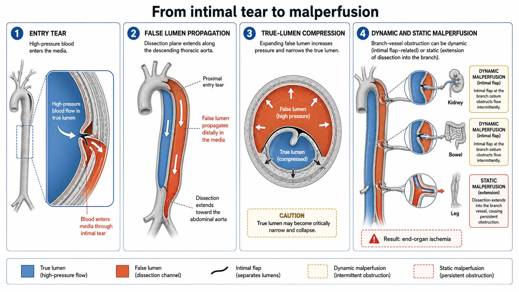
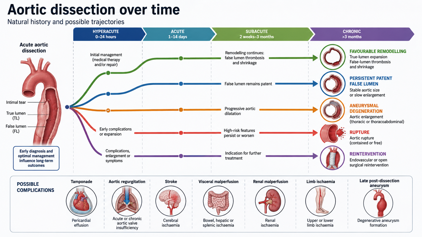
The immediate mechanical event in classical dissection is the creation of a blood-filled plane within the media. An intimal tear exposes the medial layer to pulsatile arterial pressure; blood then propagates longitudinally, often along the outer third of the media, forming a false lumen. The true lumen may become compressed, particularly in type B dissection, where dynamic obstruction can compromise visceral, renal or limb perfusion. A distal re-entry tear can partially decompress the false lumen but also permits persistent false-lumen flow. If the false lumen remains patent or partially thrombosed, late aneurysmal degeneration becomes more likely than when complete thrombosis and favourable remodelling occur. These processes explain why imaging end points such as false-lumen thrombosis, true-lumen expansion and aortic diameter stabilization are biologically plausible but must not be treated as interchangeable with all-cause survival.
The older phrase “cystic medial necrosis” is now best understood as an incomplete description rather than a universal mechanism. Medial degeneration, elastic fibre fragmentation, smooth muscle cell dysfunction and extracellular matrix abnormalities lower wall resilience, but hypertension, inherited aortopathy, bicuspid aortic valve-associated aortopathy, pregnancy, inflammatory disease, cocaine or stimulant exposure, iatrogenic injury and prior cardiac or aortic surgery can all contribute to the final event. In many acute dissections, the initiating lesion is not diffuse necrosis but focal failure of an already vulnerable aortic wall under adverse haemodynamic stress. The haemodynamic concept is clinically operationalized by anti-impulse therapy: reducing heart rate, systolic pressure and dP/dt limits propagation forces before definitive repair or during medical management.1,3
Classification: useful simplification and the need for richer anatomical language
Stanford and DeBakey systems remain indispensable because they are simple, teachable and therapeutically aligned. Stanford type A triggers emergency surgical evaluation, even when symptoms are atypical or haemodynamics are initially stable. Stanford type B requires immediate anti-impulse therapy and CTA-defined complication assessment, with endovascular repair prioritized when rupture or malperfusion is present. DeBakey classification remains useful for describing the proximal origin and distal extent of disease, particularly in surgical planning and historical literature.
Clinical presentation and diagnostic failure
The classic presentation is abrupt severe chest, back or abdominal pain that is tearing, ripping or migratory. Yet this classic description is neither sensitive nor sufficient. Patients may present with syncope, heart failure, neurological deficit, pulse deficit, limb ischaemia, abdominal pain, renal dysfunction, shock, painless malperfusion or symptoms mimicking acute coronary syndrome, pulmonary embolism, stroke, pancreatitis or renal colic. Type A dissection can produce myocardial infarction by coronary ostial involvement, and anticoagulation or thrombolysis given for misdiagnosed acute coronary syndrome can be catastrophic. Type B dissection can present as mesenteric ischaemia, acute kidney injury or limb ischaemia before back pain becomes obvious.
Diagnostic delay is one of the most modifiable causes of harm. Registries and reviews consistently show that aortic dissection is missed or diagnosed late in a substantial fraction of patients, particularly those without the stereotyped pain phenotype, those walking into the emergency department, those with normal initial chest radiography, and those whose first working diagnosis is acute coronary syndrome. The problem is not simply knowledge of a rare disease; it is failure to embed dissection into acute chest, back, abdominal and neurological diagnostic pathways.
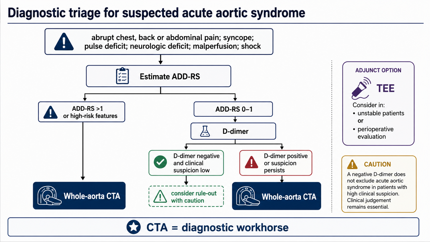
Computed tomography angiography is the practical diagnostic standard in most stable or stabilizable patients because it rapidly defines the full aorta, branch-vessel involvement, true and false lumens, entry tears, rupture signs, pericardial effusion and anatomical suitability for surgery or TEVAR. Transoesophageal echocardiography remains valuable in unstable patients, in suspected proximal dissection, and intraoperatively. MRI is accurate but less available for acute unstable triage. D-dimer has high sensitivity but poor specificity; it is best used in structured low-risk rule-out strategies rather than as an isolated test.
The ADD-RS plus D-dimer strategy attempts to formalize this logic. ADD-RS assigns risk features across high-risk conditions, high-risk pain and high-risk examination findings. In the ADvISED prospective multicentre study, combining ADD-RS with D-dimer identified low-risk groups with high sensitivity and negative predictive value, but the algorithm requires cautious use because external validation, local prevalence, assay performance, time from symptom onset and clinician gestalt all affect safety.9 A negative D-dimer should not override high-risk anatomy, high-risk symptoms or high-risk signs.
Initial management has two simultaneous objectives: prevent propagation or rupture, and maintain organ perfusion. Anti-impulse therapy usually begins with intravenous beta-blockade to reduce heart rate and shear stress, followed by vasodilators if systolic pressure remains elevated. Giving an arterial vasodilator before rate control can provoke reflex tachycardia and increase dP/dt, undermining the central physiological objective. Common targets are heart rate near or below 60 beats per minute and systolic blood pressure around 100–120 mmHg when tolerated, but targets must be individualized in shock, tamponade, severe aortic regurgitation, renal malperfusion, mesenteric ischaemia or neurological compromise.1,3
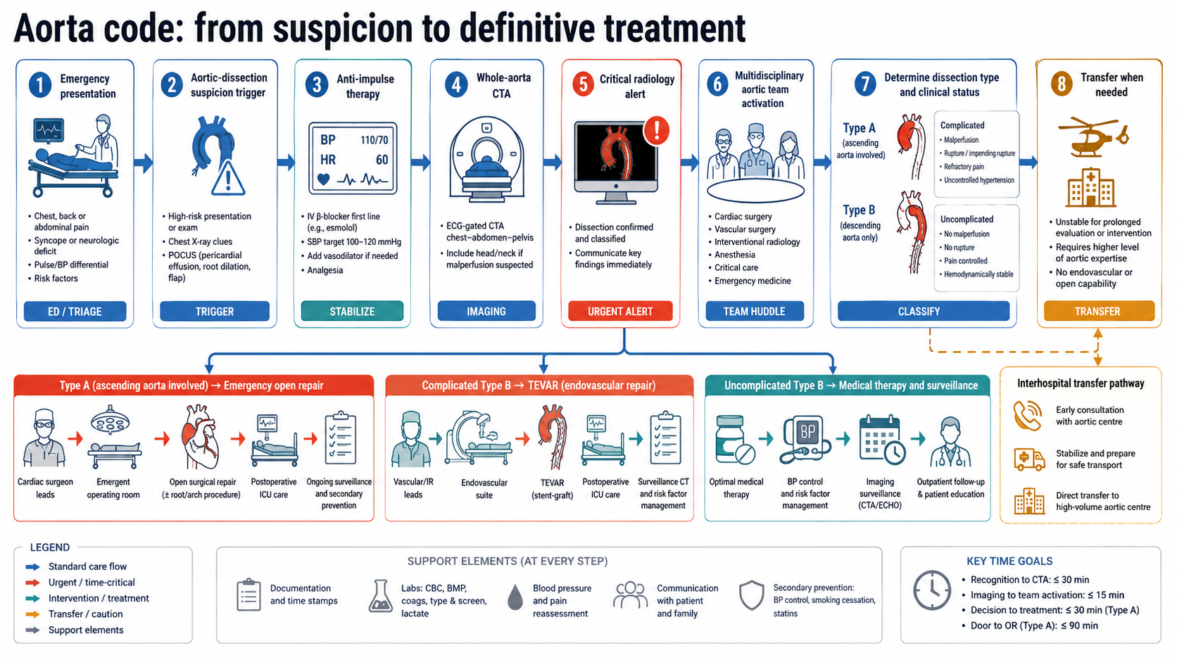
Analgesia is not a comfort measure alone; pain drives sympathetic tone and may signal ongoing propagation. Arterial-line monitoring, cross-matched blood, urgent imaging review and early involvement of cardiac surgery, vascular surgery, anaesthesia, critical care and radiology are part of treatment, not administrative steps. In high-performing systems, suspected type A dissection should activate an “aorta code” analogous to stroke or trauma pathways, because delays often occur before the patient reaches the operating room rather than after the diagnosis is made.
Type A dissection: surgical emergency and individualized proximal repair
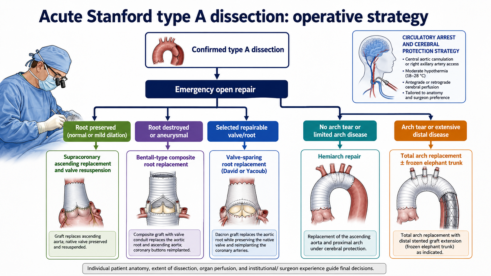
The treatment goal in acute type A dissection is to prevent death from rupture, tamponade, coronary compromise, aortic regurgitation and malperfusion by excising or excluding the proximal entry tear, replacing the ascending aorta, restoring true-lumen flow and correcting life-threatening proximal complications. Contemporary guidelines strongly support emergency operative repair for most patients with acute type A dissection, with decision-making individualized for extreme frailty, catastrophic neurological injury, prohibitive comorbidity or patient goals of care.1–3
At minimum, repair usually includes replacement of the ascending aorta and open distal anastomosis under circulatory arrest or carefully selected cerebral protection strategy. Root management depends on valve morphology, root size, intimal tear location, coronary involvement, connective tissue disease and surgical expertise. If the root is structurally preserved and the valve competent, supracoronary ascending replacement with commissural resuspension may be adequate. If the root is destroyed or aneurysmal, a composite valve graft procedure such as Bentall may be required. In selected patients, valve-sparing root replacement can avoid prosthetic valve-related complications and lifelong anticoagulation, but it is technically demanding and must not compromise emergency safety.
Contemporary data have refined but not eliminated the urgency narrative. Historical teaching often cited mortality of 1–2% per hour during the first 48 hours. More recent IRAD analyses suggest early mortality in the modern era is lower among surgically managed patients than the historical rule implies, while mortality remains very high in patients who do not undergo surgery.10 The practical implication is not to slow down; it is to replace fatalism with organized urgency. Modern imaging, transfer systems, perioperative care and high-volume aortic teams can improve outcomes, but only if the diagnosis is made and the pathway is activated.
Type B dissection: the central controversy of modern management
Type B dissection management is shaped by a tension between short-term safety and long-term aortic fate. In uncomplicated acute type B dissection, optimal medical therapy can stabilize most patients initially. The aim is strict anti-impulse therapy, pain control, surveillance imaging and long-term risk-factor control. In complicated type B dissection, TEVAR is generally preferred when anatomy is suitable because covering the proximal entry tear can restore true-lumen perfusion, reduce false-lumen pressure and treat rupture or malperfusion less invasively than open descending thoracic replacement.1–3
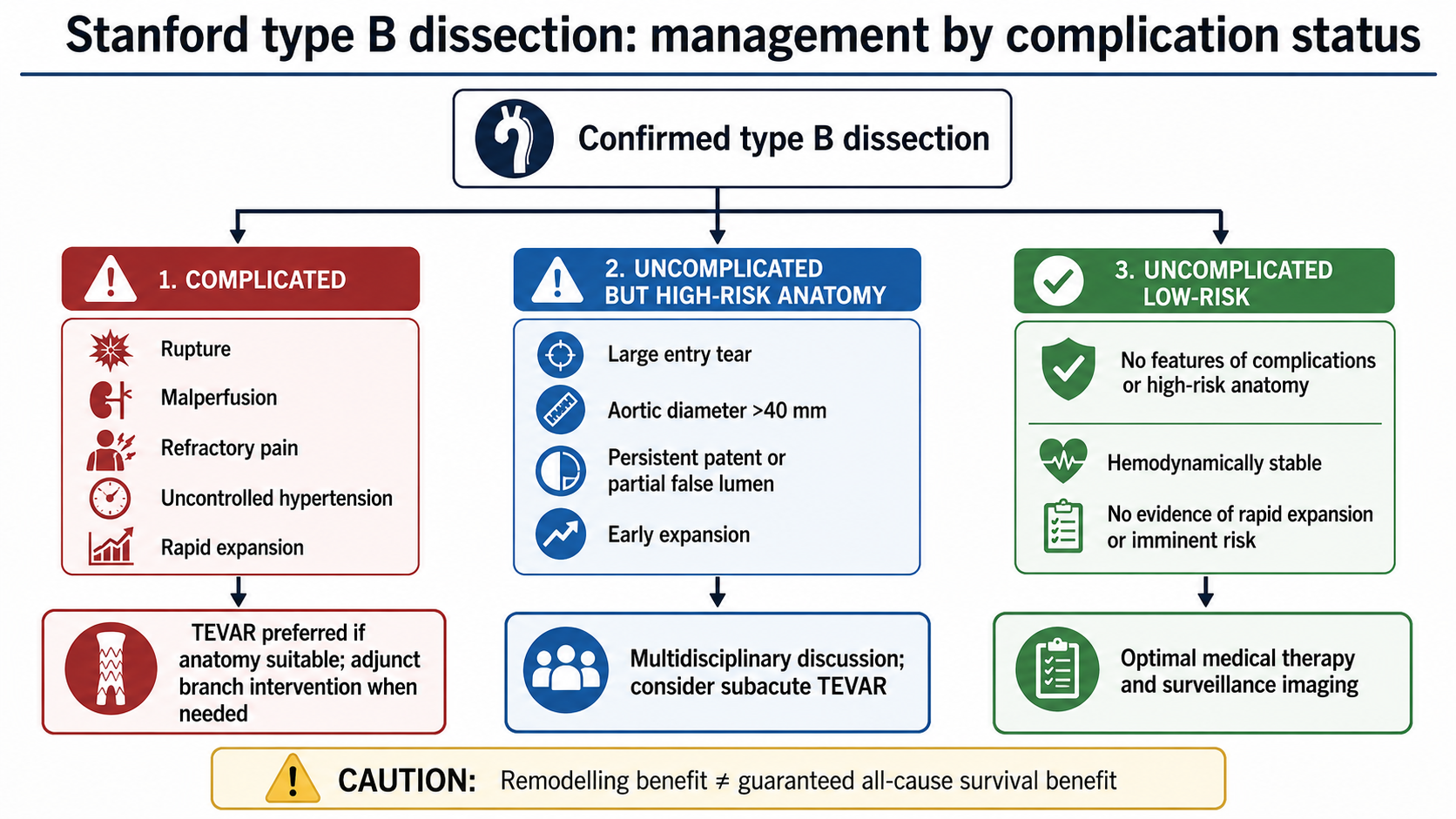
The evidence therefore supports a nuanced position: TEVAR is not merely a rescue therapy for complicated disease, but neither should it be generalized to every uncomplicated type B dissection. Imaging high-risk features—large proximal entry tear, large initial aortic diameter, rapid expansion, partial false-lumen thrombosis, persistent patent false lumen, refractory hypertension or pain, branch-vessel vulnerability and adverse morphology—should trigger multidisciplinary discussion. The subacute phase is often viewed as an attractive intervention window because the dissection membrane may be more stable than in the hyperacute period but still remodelable compared with chronic disease. Nevertheless, the decisive randomized trial for all-cause survival in well-defined high-risk uncomplicated disease remains lacking.
Chronic dissection and lifelong surveillance
Survival from the acute event does not end the disease. Chronic dissection is a dynamic state in which persistent false-lumen flow, distal re-entry, branch involvement and progressive wall degeneration can produce aneurysmal dilation years later. Patients require lifelong imaging surveillance, aggressive blood-pressure control, management of lipids and smoking, exercise counselling, and clear thresholds for reintervention. Surveillance schedules vary by guideline, operation and anatomy, but early post-discharge imaging followed by interval imaging at 3–6 months, 12 months and annually when stable is common practice.1,3
The chronic phase also reveals the consequence of initial strategy. A limited proximal repair for type A dissection may save life acutely but leave a patent distal false lumen that later requires TEVAR, branched/fenestrated repair, open thoracoabdominal replacement or hybrid intervention. Conversely, aggressive arch and frozen elephant trunk strategies may reduce later distal procedures but increase acute operative burden. In type B dissection, medical stabilization may avoid early procedural risk but can lead to late aneurysmal degeneration in a subset. Thus, initial treatment should be judged not only by in-hospital survival but by the long-term aortic trajectory.
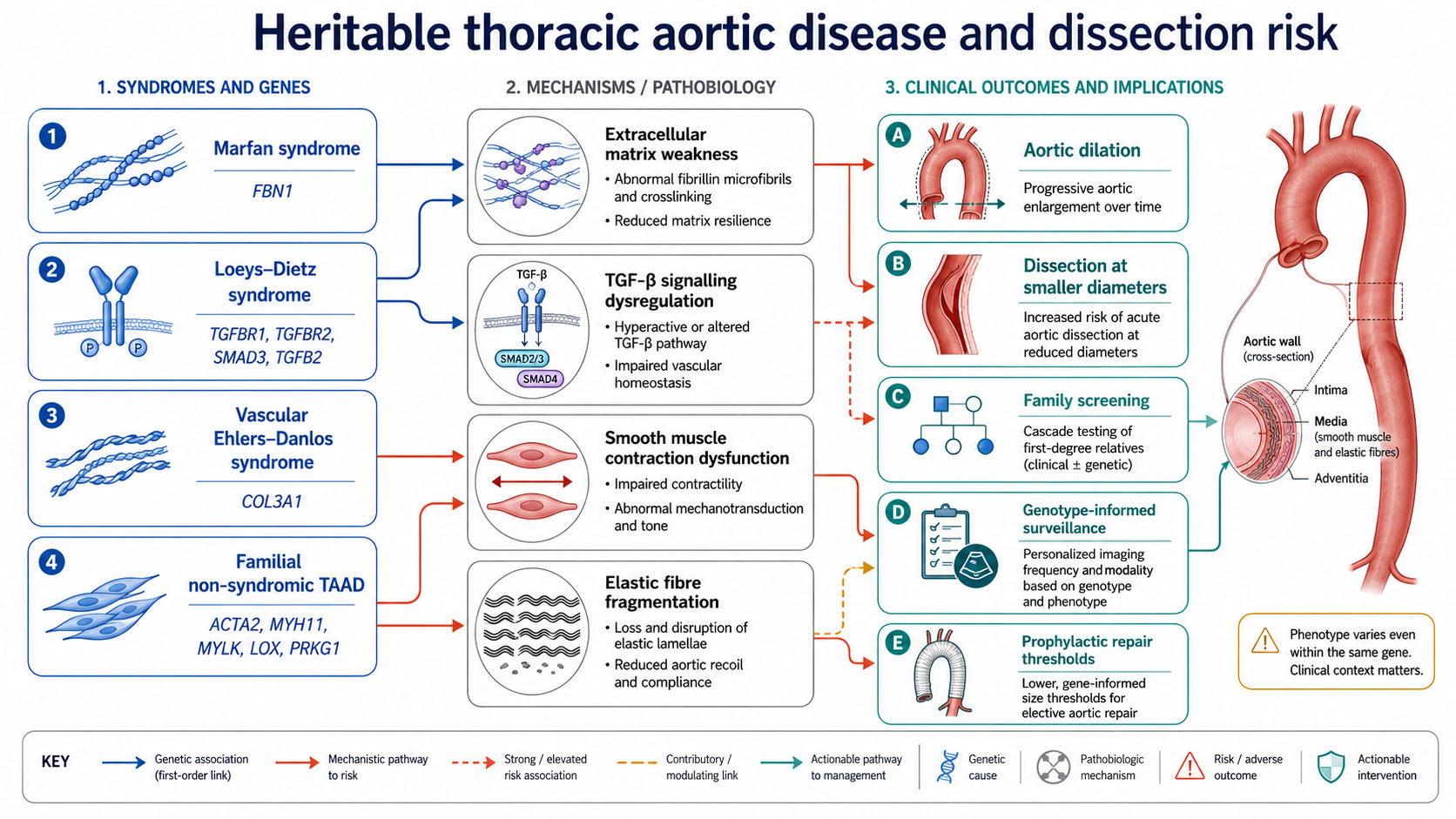
Aortic dissection in younger patients, patients with syndromic features, family history, bicuspid aortic valve, arterial tortuosity or dissection at smaller diameters should prompt consideration of heritable thoracic aortic disease. Marfan syndrome, Loeys–Dietz syndrome, vascular Ehlers–Danlos syndrome, ACTA2-related vasculopathy and other familial thoracic aortic aneurysm/dissection conditions differ in vascular distribution, dissection threshold, surgical timing and family-screening implications. Genetic diagnosis can alter thresholds for prophylactic repair, determine surveillance of the entire arterial tree and identify relatives who need imaging before symptoms occur.1,14
The conceptual shift is from diameter-only risk to genotype-informed risk. Diameter remains essential, but it does not fully capture wall biology. Some pathogenic variants dissect at smaller diameters; others carry specific risks for branch-vessel disease, early onset or aggressive progression. Genetic testing should be paired with counselling and cascade screening, because the value of identifying a pathogenic variant is realized through surveillance and prevention in relatives. For academic and educational figures, inherited aortopathy should not be presented as rare trivia but as a core pathway for preventing first catastrophic presentation.
CTA remains the central imaging modality because it converts a suspected syndrome into a map for action. The radiology report should specify Stanford/DeBakey or TEM classification, entry tear if visible, aortic diameters, arch zones, branch-vessel involvement, true and false lumen features, rupture signs, pericardial effusion, aortic valve or root implications when assessable, and prior graft or stent anatomy. For type B dissection, features relevant to TEVAR planning include proximal landing zone, left subclavian involvement, access vessels, arch curvature, iliac anatomy, false-lumen status and visceral branch perfusion.
Artificial intelligence is most plausible as a workflow accelerator rather than an autonomous diagnostic authority. Automated detection, segmentation of the aorta, true/false lumen labelling, diameter measurement, worklist prioritization and growth prediction could reduce missed diagnoses and standardize follow-up. However, AI tools must be prospectively validated across scanners, contrast protocols, motion artefacts, postoperative anatomy, intramural haematoma, penetrating ulcer and uncommon arch variants. A visually impressive model that performs well on curated retrospective CT images may still fail in the emergency department if it cannot handle incomplete scans, low contrast, artefact or low-prevalence presentations. The near-term role is human-in-the-loop triage and quantitative support.
Systems of care: aortic teams, transfer pathways and regionalization
Aortic dissection outcomes depend on system design. The best operation or stent graft is unavailable to the patient whose diagnosis is delayed, whose CTA is not protocolled correctly, whose images are not transferred, or whose receiving centre is contacted late. Guidelines increasingly emphasize multidisciplinary aortic teams and referral to experienced centres when clinically feasible.1–3 This is especially important for type A dissection, complex arch disease, malperfusion syndromes, connective tissue disease, redo aortic surgery and extensive thoracoabdominal involvement.
A practical aortic emergency pathway should define: when emergency physicians should suspect dissection; when ADD-RS and D-dimer can be used; how to obtain whole-aorta CTA rapidly; how to control heart rate and blood pressure before imaging completion; how radiology communicates critical findings; when to activate cardiac surgery and vascular surgery; how to transfer images and patients; and how to avoid harmful anticoagulation or thrombolysis when dissection remains plausible. Such pathways are a quality-improvement intervention as much as a clinical protocol.
Several tensions define the current literature. The first is the surrogate-endpoint tension in type B disease. False-lumen thrombosis and remodelling are mechanistically credible, but trials in uncomplicated type B dissection have been underpowered for all-cause mortality. The second is the aggressiveness tension in type A repair. More extensive arch and frozen elephant trunk procedures may reduce later distal aortic events but can increase early operative complexity and neurological risk. The third is the classification tension: Stanford is fast and actionable, whereas TEM and zone-based systems are more precise but require training, imaging standardization and clinical adoption. The fourth is the diagnostic tension: rule-out tools can reduce unnecessary CTA but must not create false reassurance in high-risk presentations.
Special populations remain underrepresented in randomized evidence. Pregnancy-associated dissection, elderly frail patients, connective tissue disorders, bicuspid aortopathy, iatrogenic dissection, chronic post-dissection aneurysm, non-A non-B dissection and patients in low-resource or non-aortic-centre environments are often managed by extrapolation. Inherited aortopathy trials are difficult because events are infrequent and thresholds vary by genotype. AI studies are proliferating but still need prospective workflow trials showing reduced diagnostic delay, improved outcomes or safer resource use, not merely retrospective accuracy.
Research priorities for the next five years
The most important randomized evidence gap is the optimal management of high-risk uncomplicated type B dissection. A definitive trial would need to define high-risk imaging criteria prospectively, stratify acute versus subacute intervention, require modern devices and contemporary medical therapy, and use clinically meaningful end points including all-cause mortality, aorta-related mortality, rupture, malperfusion, reintervention, spinal cord ischaemia, stroke, renal failure, quality of life and cost-effectiveness. Substudies should examine remodelling as a mediator rather than a substitute for clinical benefit.
A second priority is external validation of diagnostic pathways in diverse emergency settings. ADD-RS plus D-dimer should be tested across low- and high-prevalence populations, different D-dimer assays, variable symptom duration and real-world clinician behaviour. A third priority is imaging standardization: structured reports should link classification, measurements, branch-vessel status and treatment implications. A fourth priority is genotype-informed surveillance and prophylactic repair thresholds, especially for non-syndromic familial thoracic aortic disease.
For AI, the appropriate trial is not a leaderboard of segmentation metrics but a prospective workflow study. Useful outcomes include time from scan acquisition to critical alert, time from arrival to diagnosis, missed-dissection rate, interobserver variability in measurements, appropriateness of CTA triage, and downstream clinical outcomes. AI should also be assessed for equity, including performance across body habitus, sex, age, scanner type, institution and post-surgical anatomy.
Clinical synthesis
Aortic dissection is best understood as a time-sensitive anatomical emergency governed by three questions. Does the dissection involve the ascending aorta? Where is the entry tear and how far does the dissection extend? Is there rupture, malperfusion or high-risk morphology? These questions determine whether the patient needs emergency open repair, immediate or staged TEVAR, branch-vessel intervention, intensive medical therapy or surveillance. The most robust consensus remains straightforward: type A dissection generally requires emergency surgical repair; complicated type B dissection generally requires endovascular or surgical intervention; all patients need anti-impulse therapy and lifelong surveillance; CTA is the diagnostic workhorse; and inherited risk must be considered in younger or familial presentations.1–4
The most important intellectual caution is equally straightforward: anatomical improvement is not automatically survival benefit, and technical possibility is not universal indication. The future of the field will depend on matching treatment intensity to patient-specific risk, integrating richer classification with faster clinical workflows, and building systems that recognize dissection before irreversible malperfusion or rupture occurs.
References
- Isselbacher, E. M. et al. 2022 ACC/AHA Guideline for the Diagnosis and Management of Aortic Disease. Circulation 146, e334–e482 (2022).
- Czerny, M. et al. EACTS/STS Guidelines for diagnosing and treating acute and chronic syndromes of the aortic organ. Eur. J. Cardiothorac. Surg. 65, ezad426 (2024); also published in Ann. Thorac. Surg. 118, 5–115 (2024).
- Mazzolai, L. et al. 2024 ESC Guidelines for the management of peripheral arterial and aortic diseases. Eur. Heart J. 45, 3538–3700 (2024).
- Hagan, P. G. et al. The International Registry of Acute Aortic Dissection (IRAD): new insights into an old disease. JAMA 283, 897–903 (2000).
- Lombardi, J. V. et al. Society for Vascular Surgery and Society of Thoracic Surgeons reporting standards for type B aortic dissections. J. Vasc. Surg. 71, 723–747 (2020).
- DeBakey, M. E., Cooley, D. A. & Creech, O. Surgical considerations of dissecting aneurysm of the aorta. Ann. Surg. 142, 586–612 (1955).
- Daily, P. O., Trueblood, H. W., Stinson, E. B., Wuerflein, R. D. & Shumway, N. E. Management of acute aortic dissections. Ann. Thorac. Surg. 10, 237–247 (1970).
- Evangelista, A. et al. Insights from the International Registry of Acute Aortic Dissection: a 20-year experience of collaborative clinical research. Circulation 137, 1846–1860 (2018).
- Nazerian, P. et al. Diagnostic accuracy of the aortic dissection detection risk score plus D-dimer for acute aortic syndromes: the ADvISED prospective multicenter study. Circulation 137, 250–258 (2018).
- Harris, K. M. et al. Early mortality in type A acute aortic dissection: insights from the International Registry of Acute Aortic Dissection. JAMA Cardiol. 7, 1009–1015 (2022).
- Nienaber, C. A. et al. Randomized comparison of strategies for type B aortic dissection: the INSTEAD trial. Circulation 120, 2519–2528 (2009).
- Nienaber, C. A. et al. Endovascular repair of type B aortic dissection: long-term results of the randomized INSTEAD trial. Circ. Cardiovasc. Interv. 6, 407–416 (2013).
- Brunkwall, J. et al. Endovascular repair of acute uncomplicated aortic type B dissection promotes aortic remodelling: 1-year results of the ADSORB trial. Eur. J. Vasc. Endovasc. Surg. 48, 285–291 (2014).
- Pinard, A., Jones, G. T. & Milewicz, D. M. Genetics of thoracic and abdominal aortic diseases. Circ. Res. 124, 588–606 (2019).